An Introduction to Interchromosomal Duplications

Drosophila geneticists have used many different approaches to isolate interchromosomal duplications. The most common are outlined briefly here and numbered to match the interchromosomal duplications stock pages. For a more complete review of duplications, see chapter 15 of Drosophila: a laboratory handbook.

Go to the interchromosomal duplication stock page for the:

1. Transposition

Conceptually, the most straightforward method for making a duplication is transposition, where three chromosome breakpoints allow the movement of a segment to a new insertion site.

Transposition

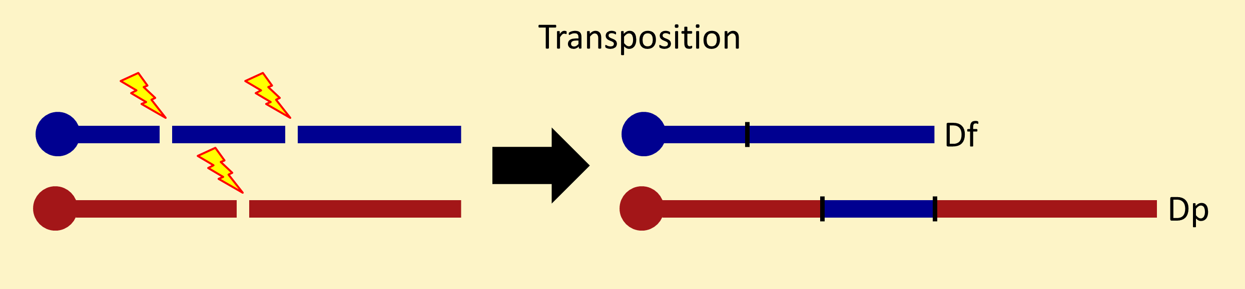
It is important to note that a transposition (Tp) is composed of a pair of chromosomes: a deletion (Df) and a duplication (Dp). The deletion and duplication chromosomes can be inherited separately, but progeny inheriting only one of these chromosomes often die from excessive aneuploidy. Transpositions are recognized in screens from one of the breakpoints disrupting a gene, from viable progeny always inheriting the Df and Dp chromosomes together (pseudolinkage), or both.

Dp and Tp symbols indicate the direction the duplicated segment moved. In a Tp(3;2) or Dp(3;2), a third chromosome segment is inserted in the second chromosome. In a Tp(2;3) or Dp(2;3), a second chromosome segment is inserted in the third chromosome.

Because transpositions are relatively rare events, geneticists have devised other methods for recovering interchromosomal duplications. 

2. Translocation

Sometimes, one of the breakpoints of a reciprocal translocation occurs near a chromosome tip, distal to vital genes. When this happens, a segment of another chromosome can be appended to the end.

Tip Swap

As long as the appended segment is not too big to cause problems from aneuploidy, the translocation segregant (one chromosome of the reciprocal translocation) can be recovered by itself and used as an interchromosomal duplication. The usefulness of this method is limited to duplicating genes near the ends of chromosomes.

3. Ring formation

If breaks occur on both sides of the centromere, a ring chromosome can be formed that carries genes near the bases of the arms.

Ring foirmation

Such chromosomes are small and exist in stocks as free duplications—extra chromosomes present in addition to the usual set of chromosomes. (Free duplications are not really “interchromosomal”, but they are used for the same experimental purposes, so we have included them here.) They are denoted by f in duplication symbols. For example, a free duplication of a third chromosome segment is denoted Dp(3;f).

4. Free duplication from large deletion

A related method is used to recover free duplications of the X chromosome. Because the centromere of the X chromosome lies at the right end, a free duplication can be formed from a large internal deletion.

Free duplication from large deletion

These chromosomes carry genes from the X tip. The presence of the yellow gene very close to the telomere allows them to be detected in screens. They can carry genes from the X base as well, but often the right breakpoint lies in basal heterochromatin proximal to genes.

5. Free duplication from deletion within an inverted X chromosome

If an X chromosome has an inverted segment, it can be used to recover a free duplication carrying genes from the middle of the X in addition to genes near the tip and base.

Free duplication from large deletion within an inverted chromosome

6. Y-linked duplication from deletion within an attached-XY chromosome

If one starts with an attached-XY chromosome, a large deletion within the X chromosome results in a Y-linked duplication of the X tip.

y-linked duplication from deletion within an attached-XY chromosone

Depending on the position of the right breakpoint, genes from the base of the X can also be present.

7. Y-linked duplication from a deletion within an attached-XY carrying an inversion

If the X chromosome of an attached-XY chromosome has an inverted segment, then a large deletion within the X can result in a Y chromosome carrying a segment from the middle of the X in addition to tip and base segments.

Y-linked duplication from a deletion within an attached-XY carrying an inversion

This is the method that was used to generate the Dp(1;Y) chromosomes in a project at the Bloomington Stock Center.

8. Y-linked duplication from two reciprocal translocations

Two sequential reciprocal translocation events can place an internal segment of any chromosome onto the Y chromosome.

 Y-linked duplication from two reciprocal translocations

Often these duplication chromosomes lack some Y chromosome genes, so they are maintained in stock with another Y chromosome.

9. Duplication from deletion within a reciprocal translocation

Deleting a segment within a reciprocal translocation can allow one of the component chromosomes to be separated from the other and act as a duplication chromosome.

Duplication from deletion within a reciprocal translocation

This approach is most useful when one translocation breakpoint lies distal to most genes.

10. Y-linked duplication from deletion within a reciprocal translocation

This method is used to place X chromosome segments on a Y chromosome. Meiotic recombination can be used to replace the normal X chromosome of an attached-XY with an X-Y translocation. Then, most of the X chromosome proximal to the translocation break can be deleted to leave a segment for the middle of the X appended to a Y chromosome.  Often a segment from the base of the X is duplicated, too.

 Y-linked duplication from deletion within a reciprocal translocation